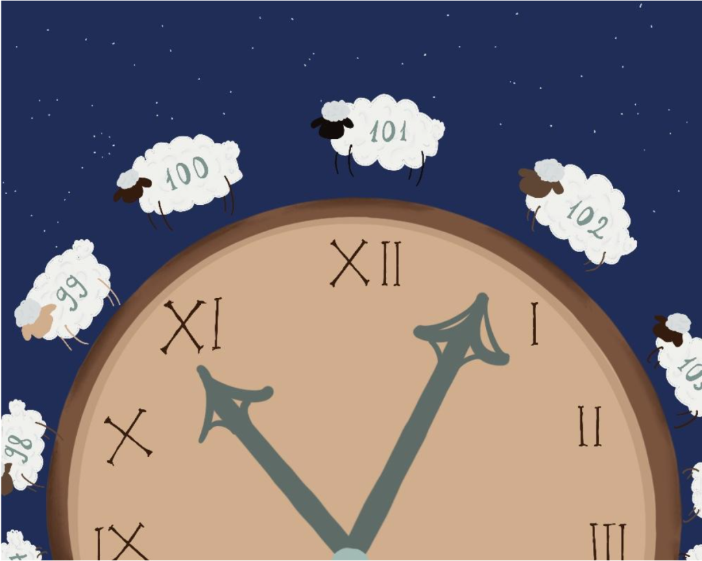
When in doubt, sleep!
Do you ever find yourself thinking that there are so many things to do, but so little time? And that in order to get everything done you need to sleep less and work more? If the answer is yes, then you might find it hard to imagine that your busy life could fit the recommended eight hours of slumber. Numerous studies show the essential role of sleep in creativity, memory and the quality of social interactions. There are many stories about groundbreaking discoveries made just after waking up. Great examples are Mendeleev’s periodic table, Kekulé’s benzene structure or Otto Loewi’s discovery of chemical neurotransmitters, which landed him a Nobel Prize. What happens if you don’t sleep Just as striking is the evidence proving the connection between not enough sleep and pathology. Sleep deprivation is associated with an overactive sympathetic nervous system and an increased level of the stress-related hormone cortisol. This leads both to a compromised immune system and to cardiovascular problems. Those include increased blood pressure and tachycardia. Over time, sleep-deprived adults become about 200% more susceptible to heart attacks or strokes than those who get enough of it. Sleep deprivation also leads to changes in the concentration of two key appetite controlling hormones: leptin and ghrelin. And because insufficient sleep is also connected to insulin resistance, it is not hard to imagine why lack of sleep is linked with type 2 diabetes and obesity. As for the neurological disorders, one of the most impactful examples is Alzheimer’s disease. Research shows that the amyloid protein – a toxin whose buildup is associated with Alzheimer’s – tends to accumulate in the middle part of the frontal lobe. This is the same region responsible for generating the electrical waves of deep NREM sleep. Comparatively, studies prove that the glymphatic system – composed of glial cells responsible for waste clearance – is most active during NREM sleep. So, most of the amyloids are supposed to be removed during this time. Without sleep, the concentration of this toxin in the brain increases. Subsequently, this increases the chance of developing Alzheimer’s. What makes for a better sleep So getting enough quality sleep is crucial. Keep in mind, sleep deprivation is considered so dangerous that the Guinness Book of World Records refuses to recognise any attempt at setting a new world record when it comes to the number of hours spent awake. Undoubtedly there are factors beyond our control that prevent us from sleeping the recommended amount of time. However, there are still some tips that could help. Those include: a consistent sleep schedule and avoiding long naps. Physical activity also helps! Oh, avoid caffeine 5-7 hours before bedtime. Not to mention, go to bed in a dark room! So maybe it is time we stopped looking at sleep as a passive waste of time. Instead let’s recognise what it actually is: an essential, indispensable part of our lives that is intertwined with every aspect of living life at its best. And at the end of the day, it seems like the answer could lie in tackling the issue the other way around: sleeping more and working less. If you found this article interesting, stay tuned and stay curious with us! You can read the full text here. See you next time. About the author… Hello! I’m Mara, a second year medical student whose interests revolve around stories, of any kind. So be it music, science, dance or graphic arts, I love looking for the story behind it. Even better if I can also have a cat around.
When in doubt, sleep! Read More »